I.) In our reflex lab, we examined 5 different parts of our body that contains reflexes. First we captured the dilation of our pupils by closing our eyes for 2 minutes, and then shining a bright light near the eye to watch the pupil decrease in size. Our eyes contain an autonomic reflex, in which the pupil allows less light to enter the eye and prevent from blindness. As the eyes respond to the extreme amount of light, they make themselves smaller in order to protect the retina and other parts of the eye from being damaged from bright light. Here is a video showing the pupil dilating and decreasing the amount of light entering the eye:
II.) We experimented with our knee-jerk reflex, also known as the Pateller reflex in the second part. We sat on a table and used a reflex hammer to hit the base of our base and initiate a kick. The thigh muscle contracts and causes the lower leg to jerk out. Knees contain the pateller reflex of jerking outward as a defensive reaction to protect from predators and provide self defensive. Furthermore, after doing 30 squats, our Pateller reflex was dulled because our muscles were tired from fatigue and the autonomic reflex that regulates our smooth muscle (upper thigh) was dulled.

III.) We examined our blink reflex by throwing a cotton ball at clear plastic wrap that covered our faces. When the ball hit the wrap, we instantly blinked out of reflex. Our eyes contain this withdrawal reflex to blink against a sudden attack in order to protect our eyes from threats as well as provide us with a moment of calmness to maintain composure and stability. By closing our eyes for a split moment, we can defy threats more effectively by channeling our fear into a split-second reflex instead of panic.
IV.) To test out plantar reflex, we traced a pen cap on the bottom of our foot starting form the heel up to the big toe. If we respond normally, our toes will clench closer together and shows that our nervous system functions properly. If someone has a Central Nervous System disorder like Multiple Scerosis, they feet would not exhibit the planter reflex because the neurons would not respond to the scrapping properly.
V.) Lastly we did an experiment with a ruler to test the speed of our response to multiple variables. Our reaction time was determined by how fast we could grab a ruler that was dropped at a random time into our hands. Here is the table showing Nicole and I's times for 3 trials:
To truly examine how our responses can be easily distracted through various external factors such as texting, we did the same trial again, except with one hand focused on texting and the other hand still trying to grab the yard stick as fast as possible. We noticed that texting slowed our reaction time to the yeard stick and increased the distance in comparison to the trails done without texting. This alludes to the importance of not texting while you drive as your reaction time is significantly slowed and can result in dangerous situations. Here is the resulting data:
In the end, I had a faster reaction time than Nicole, most likely because I had gotten more sleep than she had the previous night and I had experience from color guard to catch equipment quickly and with strength. My senses were therefore heightened and made my performance overall.
 We dissected an owl pellet that contained bone remnants of various rodents. After examining our owl pellet, our claim is that we had multiple animals in our pellet, one most prominently identified as a vole. Our first indicator that our animal was a vole is shown in the lower jaw also known as the mandible. We noticed that the teeth were primarily concentrated near the back of the jaw, and a large incisor grew out of the skull smoothly and sharpened to a point. Voles only contain a small amount of molars, that are compensated for with their large incisor. Notice in the picture to the right how the set of teeth are located near the back end of mandible and the continuity of the incisors growing out of it. Voles also contain two large incisors that come out of the skull, The attachment area of the mandible contains 3 rungs that protrude in a very distinct shape with the middle edge also known as the (condyloid process) pointing upwards and the lower point curving downward. (The pictures below are from Brandon and William´s pellet). Both the vole skull and human skull have a mandible and maxilla with molars that retreat between them. Teeth are placed in a striaght set within the mandible allowing for more detailed chewing. They both also contain a condyloid process which attaches the jaw to the skull. This knob along with the contraction of the temporalis muscle allows the jaw to move. The vole however contains two large incisors that protrude outside of the jaw, allowing for enhanced accuracy of capturing prey. The human incisors are located inside of the mouth, succinct with the rest of the teeth.
 Compared to the other rodent skulls, the vole's skull has a large and hollow indent down the center. There are also large sockets for the eyes and the points on the side are most likely the zygomatic arch that has been cut in half. In the picture below, you can clearly see the space where the 2 large incisors were originally located. Both the human skull and vole skull contain a zygomatic arch that is superior to the mandible bone, and distinct inversions of the skull where the eye sockets belong.
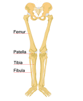
 Another distinct trait we noticed to conclude our observation of a vole is the straight and simplicity of the upper leg limbs. While other animals contained bones that were shorter or have more of a cuboidal structure, the ulna and radius of a vole are long and skinny, similar to the anatomy of a human femur shown on the picture to the right, the vole´s humerous also has a small bump that sticks out of the bone, making it clear to indicate it belongs to a vole also making it relatable to the femur in a human body. Both the vole and human femurs are long bones with the proximal and distal epiphysis located at two ends. While the human femur contains a smooth structure all the way down the limb, the vole's limb contains the small ridge protruding out.


