 We dissected an owl pellet that contained bone remnants of various rodents. After examining our owl pellet, our claim is that we had multiple animals in our pellet, one most prominently identified as a vole. Our first indicator that our animal was a vole is shown in the lower jaw also known as the mandible. We noticed that the teeth were primarily concentrated near the back of the jaw, and a large incisor grew out of the skull smoothly and sharpened to a point. Voles only contain a small amount of molars, that are compensated for with their large incisor. Notice in the picture to the right how the set of teeth are located near the back end of mandible and the continuity of the incisors growing out of it. Voles also contain two large incisors that come out of the skull, The attachment area of the mandible contains 3 rungs that protrude in a very distinct shape with the middle edge also known as the (condyloid process) pointing upwards and the lower point curving downward. (The pictures below are from Brandon and William´s pellet). Both the vole skull and human skull have a mandible and maxilla with molars that retreat between them. Teeth are placed in a striaght set within the mandible allowing for more detailed chewing. They both also contain a condyloid process which attaches the jaw to the skull. This knob along with the contraction of the temporalis muscle allows the jaw to move. The vole however contains two large incisors that protrude outside of the jaw, allowing for enhanced accuracy of capturing prey. The human incisors are located inside of the mouth, succinct with the rest of the teeth.
 Compared to the other rodent skulls, the vole's skull has a large and hollow indent down the center. There are also large sockets for the eyes and the points on the side are most likely the zygomatic arch that has been cut in half. In the picture below, you can clearly see the space where the 2 large incisors were originally located. Both the human skull and vole skull contain a zygomatic arch that is superior to the mandible bone, and distinct inversions of the skull where the eye sockets belong.
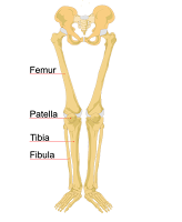
 Another distinct trait we noticed to conclude our observation of a vole is the straight and simplicity of the upper leg limbs. While other animals contained bones that were shorter or have more of a cuboidal structure, the ulna and radius of a vole are long and skinny, similar to the anatomy of a human femur shown on the picture to the right, the vole´s humerous also has a small bump that sticks out of the bone, making it clear to indicate it belongs to a vole also making it relatable to the femur in a human body. Both the vole and human femurs are long bones with the proximal and distal epiphysis located at two ends. While the human femur contains a smooth structure all the way down the limb, the vole's limb contains the small ridge protruding out.
 |
| Measurements for the most prominent rodent in pellet (Vole) |

No comments:
Post a Comment